Question 1
1. Which layer(s) of the abdominal wall is (are) composed of loose connective tissue with a variable amount of fat? Choose all that apply.
Choices:
- extraperitoneal fascia
- superficial (Camper’s) fascia
- parietal peritoneum
- transversalis fascia
- deep (Scarpa’s) fascia
Answers:
- extraperitoneal fascia
- superficial (Camper’s) fascia
Question 2
2. Which layer of the abdominal wall forms its internal surface?
Choices:
- superficial (Camper’s) fascia
- deep (Scarpa’s) fascia
- transversalis fascia
- extraperitoneal fascia
- parietal peritoneum
Answers:
Question 3
3. What is the name of the connective tissue structure that encloses the one vertically-oriented muscle of abdomen?
Choices:
- rectus sheath
- linea alba
- rectus aponeurosis
- linea rectus
Answers:
Question 4
4. Which skeletal muscle(s) of the abdominal wall is (are), in part, aponeurotic? Choose all that apply.
Choices:
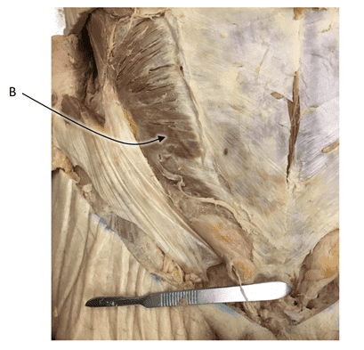
- transversus abdominis
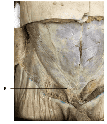
- external oblique
- internal oblique
- rectus abdominis
Answers:
- transversus abdominis
- external oblique
- internal oblique
Question 5
5. Place the three flat muscles of the abdominal wall in order, from superficial to deep.
Choices:
- external oblique
- internal oblique
- transversus abdominis
Answers:
- external oblique
- internal oblique
- transversus abdominis
Question 6
6. Which muscle layer of the anterolateral abdominal wall has fibres oriented in a superomedial direction?
Choices:
- internal oblique
- external oblique
- transversus abdominis
- rectus abdominis
Answers:
Question 7
7. The connective tissue structures that separate the subdivisions of the rectus abdominis are the ________________ ____________________.
Choices:
Answers:
Question 8
8. The landmark that demarcates the lateral border of the rectus abdominis is the linea ___________.
Choices:
Answers:
Question 9
9. Above the arcuate line, the aponeuroses of which muscles contribute to the anterior lamina of the rectus sheath? Choose all that apply.
Choices:
- external oblique
- internal oblique
- none of the above
- transversus abdominis
Answers:
- external oblique
- internal oblique
Question 10
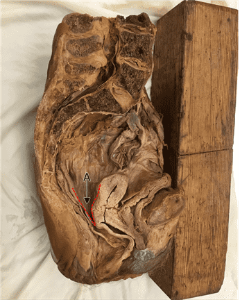
10. Below the arcuate line, with which layer of the abdominal wall is the posterior surface of rectus abdominis in contact?
Choices:
- superficial (Camper’s) fascia
- deep (Scarpa’s) fascia
- transversalis fascia
- extraperitoneal fascia
- parietal peritoneum
Answers:
Question 11
11. At what level is the arcuate line located?
Choices:
- at the umbilicus
- midway between the umbilicus and the costal margin
- midway between the umbilicus and the pubic crest
- at the costal margin
- at the pubic crest
Answers:
- midway between the umbilicus and the pubic crest
Question 12
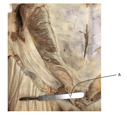
12. The aponeuroses of which muscles contribute to the formation of the conjoint tendon?
Choices:
- transversus abdominis
- internal oblique
- none of the above
- external oblique
Answers:
- transversus abdominis
- internal oblique
Question 13
13. Which intercostal nerves contribute to the innervation of the abdominal wall?
Choices:
- T9 - T11
- T7 - T11
- T5 - T11
- T3 - T11
- T1 - T11
Answers:
Question 14
14. What functional fibre types are present in the iliohypogastric and ilioinguinal nerves? Choose all that apply.
Choices:
- sympathetic postganglionic
- sensory
- somatic motor
- parasympathetic preganglionic
- parasympathetic postganglionic
- sympathetic preganglionic
Answers:
- sympathetic postganglionic
- sensory
- somatic motor
Question 15
15. What functional fibre types are present in cutaneous nerves? Choose all that apply.
Choices:
- sympathetic postganglionic
- sensory
- parasympathetic preganglionic
- parasympathetic postganglionic
- sympathetic preganglionic
- somatic motor
Answers:
- sympathetic postganglionic
- sensory
Question 16
16. Lymph from the upper quadrants of the abdominal wall primarily drains into the right and left _____________ lymph nodes.
Choices:
Answers:
Question 17
17. Lymph from the lower quadrants of the abdominal wall primarily drains into the right and left _____________ ______________ lymph nodes.
Choices:
Answers:
Question 18
18. In the fetus, the tubular evagination of the peritoneal cavity that forms alongside the gubernaculum during the descent of the testes is the ____________ _______________.
Choices:
Answers:
Question 19
19. The ______________ _______________ is the combined testicular vessels, nerves, lymphatics and the vas deferens.
Choices:
Answers:
Question 20
20. The passage through the abdominal wall formed by the processus vaginalis, and ultimately, through which the testes pass to gain access to the scrotum is the _____________ _________________.
Choices:
Answers:
Question 21
21. In the developed male, the _________ _________ occupies the inguinal canal.
Choices:
Answers:
Question 22
22. The isolated sack of serous membrane, derived from the processus vaginalis, that largely surrounds the testes in the developed male is the ________________ __________________ _________________.
Choices:
Answers:
Question 23
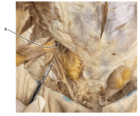
23. The deep inguinal ring is located halfway between the ASIS and pubic tubercle, immediately ____________ to the inguinal ligament.
Choices:
Answers:
Question 24
24. Lymph from the scrotum drains into the __________ _______ nodes.
Choices:
- superficial inguinal
- inguinal superficial
Answers:
- superficial inguinal
- inguinal superficial
Question 25
25. Lymph from the testes drains into the __________ nodes.
Choices:
- para-aortic
- paraaortic
- para aortic
Answers:
- para-aortic
- paraaortic
- para aortic
Question 26
26. What term describes a loop of bowel that has become fixed within a hernia?
Choices:
Answers:
Question 27
27. A patent processus vaginalis is the anatomical basis of a(n) _______________ inguinal hernia.
Choices:
Answers:
Question 28
28. An indirect inguinal hernia is located _______________ to the inferior epigastric vessels.
Choices:
Answers:
Question 29
29. A(n) ______________ inguinal hernia has a congenital basis.
Choices:
Answers:
Question 30
30. A direct inguinal hernia is located _______________ to the inferior epigastric vessels.
Choices:
Answers:
Question 31
31. A direct inguinal hernia gains access to the superficial fascia and scrotum by passing through the ____________ ____________ ring.
Choices:
Answers:
Question 32
32. A(n) ___________ inguinal hernia is described as being acquired.
Choices:
Answers:
Question 33
33. Structures that attach visceral organs to the internal surface of the body wall are called __________.
Choices:
Answers:
Question 34
34. The position of viscera that are suspended within the peritoneal cavity by mesenteries is described as ______________.
Choices:
Answers:
Question 35
35. The position of viscera that are embedded in the extraperitoneal fascia of the posterior abdominal wall are said to be _________________.
Choices:
Answers:
Question 36
36. The peritoneum that covers an abdominal organ is analogous to the _______________ that covers the heart and the ______________ that covers the lungs. (separate answers with one space)
Choices:
Answers:
Question 37
37. Peritoneal fluid is secreted by the ______________.
Choices:
Answers:
Question 38
38. The serous membrane that lines the internal surface of the abdominopelvic cavity is called ____________ ____________.
Choices:
Answers:
Question 39
39. The serous membrane that covers intraperitoneal organs is called ____________ ____________.
Choices:
Answers:
Question 40
40. The position of an abdominal organ that has a mesentery in the fetus, but during development becomes fixed to the posterior body wall, is said to be ___________ ______________.
Choices:
- secondarily retroperitoneal
Answers:
- secondarily retroperitoneal
Question 41
41. Which organs develop, even in part, within the ventral mesentery? Choose all that apply.
Choices:
- the pancreas
- the gall bladder
- the liver
- the spleen
Answers:
- the pancreas
- the gall bladder
- the liver
Question 42
42. The two subdivisions of the peritoneal cavity are the _______________ sac and the ____________ sac. (separate answers with one space)
Choices:
- greater lesser
- lesser greater
Answers:
- greater lesser
- lesser greater
Question 43
43. The greater and lesser sacs are connected via the _________ foramen.
Choices:
Answers:
Question 44
44. Inflammation of peritoneum causes an achy, poorly-localized pain that comes and goes in waves. How would you describe this peritoneum? Choose all that apply.
Choices:
- innervated by sensory fibres in autonomic nerves
- visceral peritoneum
- covering an abdominal organ
- innervated by sensory fibres in somatic nerves of the body wall
- parietal peritoneum
- on the internal surface of the body wall
Answers:
- innervated by sensory fibres in autonomic nerves
- visceral peritoneum
- covering an abdominal organ

